This article has been reviewed according to Science X's editorial process and policies. Editors have highlighted the following attributes while ensuring the content's credibility:
fact-checked
peer-reviewed publication
trusted source
proofread
Noninvasive, continuous fluorescence monitoring of bilirubin photodegradation

Skoltech researchers have refined a technique used in bilirubin blood tests for diagnosing newborn jaundice and fine-tuning phototherapy prescribed for it. Jaundice affects up to 80% of preterm infants, who are treated with blue light phototherapy and conventional drugs.
However, so far no standard scientifically grounded guidelines are available specifying the precise color of light, irradiation power and duration. These parameters need to be optimized, considering that phototherapy is not free of side effects. To enable this, scientists require better ways to measure bilirubin levels, which is exactly what the new study in Physical Chemistry Chemical Physics has contributed to.
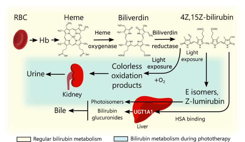
In as many as 8 in 10 preterm babies, the liver is unable to cope with bilirubin—a toxic product of red blood cell decay. This leads to elevated bilirubin levels in the blood and the tell-tale yellowish skin color.
Newborn jaundice therapy usually involves exposing the baby to blue light. This transforms the bilirubin molecules so that they can be eliminated by the body bypassing the biochemical reaction that is disrupted in a sick infant's liver.
However, there are no standard guidelines for how long blue light exposure should last, what the optimal irradiation power is, and how it all relates to disease severity.
To resolve this uncertainty, researchers need a way to measure bilirubin concentrations in the tissue more accurately than is now possible. Skoltech scientists suggest supplementing the currently used technique, which detects how much of the incident light is absorbed by the solution, with fluorescence measurements.
Fluorescence refers to molecules reemitting absorbed light at a lower frequency. That is, the light gets redder and carries less energy. (The lost energy mostly goes toward heating or configuration changes, although other mechanisms are involved, too.) Having measured fluorescence for a range of known bilirubin concentrations, you can then use these data to infer concentrations from observed fluorescence. This is intended to supplement the existing approach for greater accuracy, not to replace it.

"We took liquid samples of bilirubin at different concentrations, spanning both normal and pathological levels, illuminated them with blue light, and monitored fluorescence. The most light was emitted at a wavelength of 532 nanometers, which is green," the lead author of the study, Sergei Perkov of Skoltech Photonics, commented.
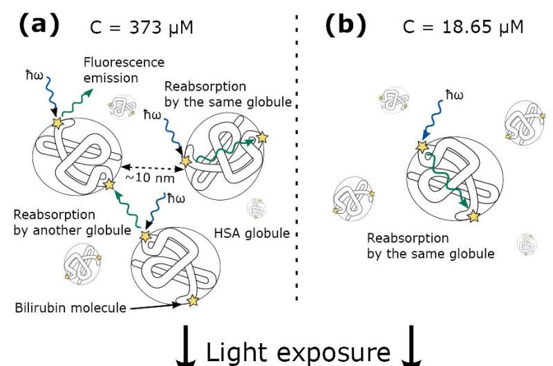
Now, it is to be expected that for a high-concentration solution, fluorescence will increase as you maintain blue light exposure. The reason is that in a concentrated solution, there is so much bilirubin all over the place that much of the fluorescent light gets reabsorbed by other bilirubin molecules before it can be detected.
As the concentration gradually goes down under blue light illumination, this so-called quenching becomes less pronounced and more fluorescence should be—and was—observed. So far, so good. But surprisingly, a similar trend presented itself for a low-concentration solution, too.

"Our current hypothesis is this," Perkov explained, remarking that at this point, this is merely a conjecture. "We know that bilirubin is transported by a protein called albumin, each molecule of which has multiple 'slots' available for it. And no matter what the concentration is, some of the fluorescence might be quenched by the neighboring bilirubin molecules carried together on one albumin. So even if the albumin molecules in the low-concentration solution are too far apart for the regular fluorescence quenching to happen, this other mechanism works regardless of concentration, and we think this explains our unexpected findings."
The scientists have thus highlighted an unexpected effect in fluorescence dynamics and supplied a calibration plot tying observed fluorescence to bilirubin concentrations. The team hopes that combined with the existing measurement technique, fluorescence monitoring will eventually make phototherapy of neonatal jaundice safer, more effective, and better tailored to individual patients.
More information: Sergei Perkov et al, Noninvasive, continuous fluorescence monitoring of bilirubin photodegradation, Physical Chemistry Chemical Physics (2023). DOI: 10.1039/D2CP03733E
Journal information: Physical Chemistry Chemical Physics
Provided by Skolkovo Institute of Science and Technology


